In these cases there may be follow-up imaging performed and comparison to past studies to see if there has been enlargement in the interim. And lower paraaortic region 11 mm.
 Thorax Usmle Anatomy Google Search Lymph Nodes Lymphatic System Lymph Massage
On imaging meaning they are slightly larger than usual but not necessarily a cause for concern.

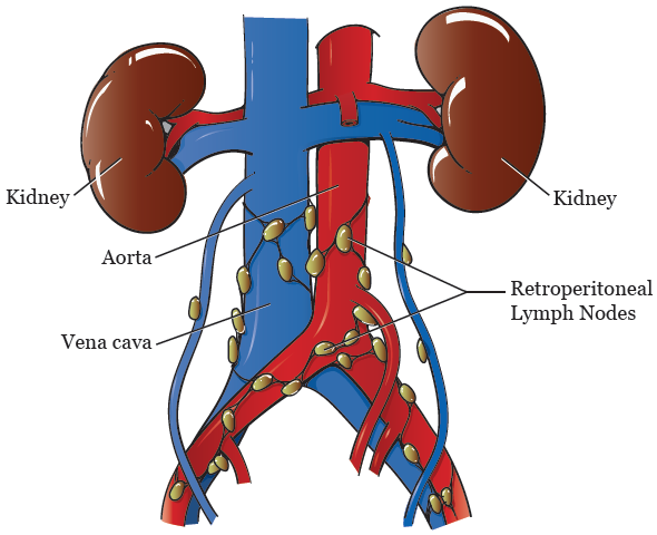
Normal para aortic lymph node size. Para-aortic lymph nodes often shortened to para-aortic nodes are part of the retroperitoneal nodes and are located anterior to the left lumbar trunk 1 and above and below the left renal vein prior to the flow of lymph into the cisterna chyli 2-4. When the axial ratio of a lymph node was greater than 05 07 10 and the maximum diameter of the long or short axis was greater than 12 mm 10 mm 8 mm or 6 mm the node was considered metastatic. Abnormal nodes are also seen as soft-tissue densities but of greater size and number.
Right superclavicular node classic sign of intrathoracic. In July the scan showed 2 iliac lymph nodes as enlarged which makes sense bc it sounds like the ovary drains into those nodes and I had just had the surgery. Identify salivary glands by location as non-lymph nodes.
Supraclavicular fossa most significant area. Retrocrural space 6 mm. However there are sooooo many other.
Porta hepatis 7 mm. Left supraclavicular node Virchows node classical sign of abdominal process. 1052018 What size of lymph node in neck consider not normal in 40 years old women.
12292002 For the most part lymph nodes greater than 1 cm are more worrisome than lymph nodes less than 1 cm. 2102021 The paraaortic lymph node is one of several masses of lymph tissue located near the aorta right in front of several lumbar vertebrae. We aimed to assess the prognostic role of visible para-aortic lymph nodes PALNs.
When lymph nodes were greater than 12 mm 10 mm 8 mm or 6 mm in longo or short-axis diameter the nodes were considered metastatic. As part of the lymphatic system these nodes help drain. These lymph nodes are referred to.
Visible para-aortic lymph nodes of 2 mm in size are common metastatic patterns of colorectal cancer CRC seen on imaging. 2 Shape and size. Often indicates a process deep in body.
Portacaval space 10 mm. Upper paraaortic region 9 mm. Upper paraaortic region 9 mm.
And lower paraaortic region 11 mm. Portacaval space 10 mm. Normal retroperitoneal lymph nodes are either invisible or appear on CT as small round or oval densities less than 1 cm in diameter Fig.
The upper limit for the diameter is not the same for all lymph nodes but varies with the position of the node in the para-aortic chains ranging from 7 to 15 mm with increasing diameters in the caudal direction. Lower paraaortic lymph nodes larger than 11 mm by short-axis measurement are abnormal. Another term for one is a periaortic lymph node.
Sometimes lymph nodes are borderline-enlarged. The upper limits of normal by location were as follows. With use of a short-axis node diameter of 1 cm as the upper limit of node size CT will detect mediastinal lymph node enlargement in about 60 of patients with node metastases CT sensitivity whereas about 70 of patients with normal nodes will be classified as normal on CT CT specificity.
Retrocrural space 6 mm. 2 doctor answers 2 doctors weighed in 90000 US. Their prognostic value however remains inconclusive.
Paracardiac 8 mm. And lymph nodes greater than 2 cm are even more worrisome. Adenopathy can occasionally be confluent or calcified.
Doctors in 147 specialties are here to answer your questions or offer you advice prescriptions and more. 3262020 The para-aortic lymph nodes are located above the left renal vein near the aorta and lumbar vertebrae. Gastrohepatic ligament 8 mm.
Dr Daniel J Bell and Dr Mila Dimitrijevic et al. Porta hepatis 7 mm. Gastrohepatic ligament 8 mm.
What is the normal size of a lymph node in the abdomen. Usually around 1 cm is the normal size but I think my husband had some in his axillary area if I remember right that were a little bigger but they called them reactive as in infection or inflamation and not related to cancer. The upper limits of normal by location were as follows.
Identify carotid arterybulb by pulsation as non-lymph nodes. So then I had a CT scan yesterday which is showing that those 2 nodes have decreased in size which is Great but now I have an enlarged para-aortic lymph node. My doctor was unconcerned.
With the aim of obtaining a normal material the diameters of the lymph nodes were measured on lymphograms that had been considered to be normal from 95 patients. There is a left and right para-aortic lymph node and they both work to process drainage from the upper thorax and abdominal cavity.
 Lymph Drainage Of The Cervix Uteri Is Complex Bilateral And Can Download Scientific Diagram
 The Iaslc Lymph Node Map Including The Proposed Grouping Of Lymph Node Download Scientific Diagram
 About Your Retroperitoneal Lymph Node Dissection Memorial Sloan Kettering Cancer Center
:background_color(FFFFFF):format(jpeg)/images/library/7344/oBUrVH5274pkjRv8PvG7g_Celiac_lymph_nodes_01.png) Retroperitoneal Space And Associated Lymph Nodes Kenhub
Retroperitoneal Space And Associated Lymph Nodes Kenhub
 Common Iliac Lymph Nodes An Overview Sciencedirect Topics
What Illnesses Are Associated With Upper Cervical Lymph Nodes 2b And 3 Quora
 Department Of Surgery Lymphadenectomy
 Common Iliac Lymph Nodes An Overview Sciencedirect Topics
 Schematic Shows The Seven Subgroups Of Paraaortic Lymph Nodes The Download Scientific Diagram
 Mediastinal Lymph Node Levels Used To Compare The Results Of Pet Ct Download Scientific Diagram
 Lymph Node Regions Illustration Radiology Case Radiopaedia Org
 Lymph Node Regions Axillary Lymph Nodes Wikipedia Lymph Nodes Lymph Massage Lymphatic System
 Classification Of Pelvic And Paraaortic Lymph Nodes 1 Paraaortic Download Scientific Diagram
 Schematic Shows The Major Pelvic And Retroperitoneal Lymph Node Groups Download Scientific Diagram
 Lymph Node Size At Various Anatomic Sites Short Axis Diameter Upper Download Table
 Living Without Lymph Nodes Ot Zone Inc
 Common Iliac Lymph Node Group Illustration A And Axial Download Scientific Diagram

